

Our Scientific Foundation
Mitochondria are dynamic organelles whose morphology is intimately linked to energy metabolism, integrity checks, and stress responses. A delicate balance of fission and fusion events dictates cell fate, determining whether damaged mitochondria are recycled (mitophagy) or entire cells are eliminated (apoptosis).
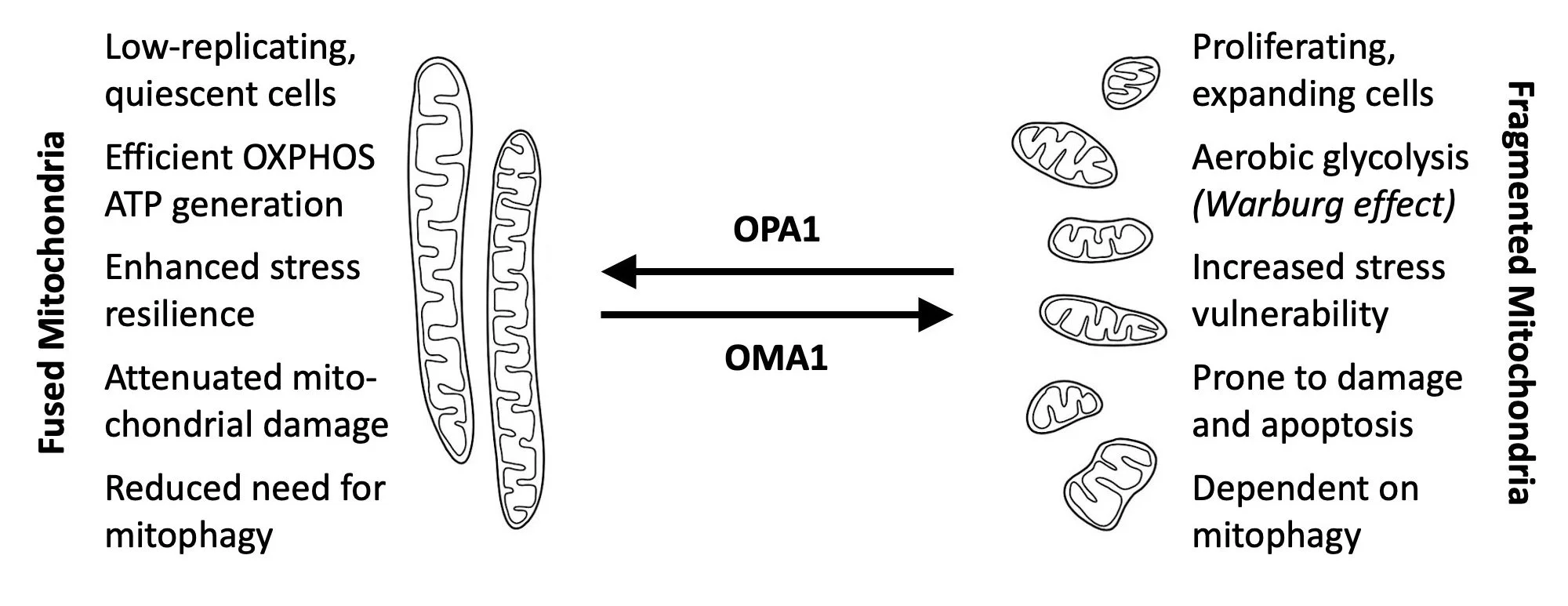

OMA1: The Mitochondrial Master Regulator
Often described as a "master regulator" of mitochondrial health, OMA1 is a multifunctional protease embedded in the inner mitochondrial membrane. Acting as a critical sensory hub, it monitors cellular stress and orchestrates a variety of life-or-death responses.
Key Functions and Mechanisms
Morphology & Cell Fate: In response to stress, OMA1 cleaves OPA1, a protein responsible for maintaining mitochondrial membrane structure. This cleavage is a hallmark event that can trigger mitophagy (the recycling of damaged mitochondria) or initiate apoptosis (programmed cell death).
The Integrated Stress Response (ISR): OMA1 cleaves DELE1, which signals the cytosol to stall general protein synthesis while prioritizing the production of protective chaperones to combat stress.
Quality Control & Dynamics: In coordination with the protease PARL, OMA1 regulates mitochondrial fission through PGAM5 and clears misfolded or misrouted proteins, such as PINK1, ensuring the inner membrane remains functional.
Proteolytic Balance: Its activity is finely tuned through reciprocal hydrolysis with the i-AAA protease, a feedback loop that balances mitochondrial capacity with the necessity of cell death signaling.
The Luke-S1—a Powerful Drug Discovery Engine
712 North's core technology is the Luke-S1 assay, a powerful, proprietary, high-throughput drug screening assay. This bioluminescence reporter is designed to screen for small molecule modulators of OMA1 protease activity. Because the assay's signal is inversely proportional to OMA1’s activity, it is uniquely efficient at identifying both inhibitors (potential treatments for neurodegenerative or cardiovascular disorders) and activators (potential treatments for cancer).